Upper Leg Tendon Anatomy - Tushank Artworks - Page 2 : The pads of the machine are situated at the achilles tendon.. Originates from the lateral condyle of the tibia and the medial surface of the fibula. The peroneus longus originates at the head of your fibula and the upper half of the shaft of your fibula on the outer part of your lower leg. This may result in tendon subluxation; Tendons are fibrous cords attached to muscles and bone. Hands are outstretched, holding onto the handles of the bench.
Leg muscle anatomy chart | amulette. The human leg, in the general word sense, is the entire lower limb of the human body, including the foot, thigh and even the hip or gluteal region. In this upper leg tutorial, i go over all the major points of the upper leg to take your sculpting skills to the next level. Hands are outstretched, holding onto the handles of the bench. Related posts of muscle anatomy upper leg.
Diagram Of Ligaments And Tendons Under Arm / Anatomy Of ... from i.pinimg.com Horse leg basic anatomy tendons подробнее. How does achilles tendon rupture occur… why are achilles piercings dangerous? .16 penile numbness and perineum tenderness.18 any suggested exercises or stretches?.22 leg musculature 209 elbow tendonitis and saddle sores. Study upper leg anatomy flashcards from tony hao's university of leicester class online, or in brainscape's iphone or android app. Human forearm anatomy inside arm anatomy upper arm anatomy skin left lower arm anatomy leg muscle and tendon anatomy arm anatomy names posterior thigh tendon anatomy feet tendon anatomy leg tendon anatomy shoulder tendon anatomy foot tendon anatomy hip. It is formed when the soleus muscle tendon joins with the gastrocnemius tendon. Plantar flexion of the foot, ankle joint stabilizer. This article will discuss the anatomy and function of the achilles tendon.
Concept conceptual 3d illustration fit strong back upper leg human anatomy, anatomical muscle isolated white background for body medical health tendon foot and biological gym fitness muscular system.
The image is available for download in high resolution quality up to 2938x2938. The tendons that control movement in your hands, wrists and fingers run through your forearm. In this upper leg tutorial, i go over all the major points of the upper leg to take your sculpting skills to the next level. Study upper leg anatomy flashcards from tony hao's university of leicester class online, or in brainscape's iphone or android app. Tendons are situated between bone and muscles and are bright white in colour. Related posts of muscle anatomy upper leg. Current techniques have tended to anatomical reconstruction of the lcl, pt and pf. Hands are outstretched, holding onto the handles of the bench. Also, i give a sculpting lecture in zbrush and timelapse video to show how i build the major shapes. Localized anatomy of the hamstring muscles including semimembranosus, semitendinosus, biceps the hamstrings refer to 3 long posterior leg muscles, the biceps femoris, semitendinosus, and semimembranosus. Leg anatomy muscles and tendons how to fix achilles. There are four muscles in the anterior compartment of the leg. Common tendon of superficial posterior leg muscles;
The thigh bone, or femur, is the large upper leg bone that connects the lower leg bones (knee joint) to the pelvic bone (hip joint). Tendon, tissue that attaches a muscle to other body parts, usually bones. Lateral (fibular) collateral ligament (fcl) upper part middle part lower part popliteus tendon (pt) upper part i. Collectively, they act to dorsiflex and invert the foot at the ankle joint. Thompson's test, achilles tendon rupture.
Appendicular Muscles of the Pelvic Girdle and Lower Limbs ... from pressbooks-dev.oer.hawaii.edu Tendon, tissue that attaches a muscle to other body parts, usually bones. Current techniques have tended to anatomical reconstruction of the lcl, pt and pf. The lower leg is comprised of two bones, the tibia and the smaller fibula. An anatomical and biomechanical study. Lie prone on a hamstring curl machine. The thigh bone, or femur, is the large upper leg bone that connects the lower leg bones (knee joint) to the pelvic bone (hip joint). Hands are outstretched, holding onto the handles of the bench. Leg anatomy muscles and tendons how to fix achilles.
You can read more about wrist tendons and the anatomy of the upper extremity, and view anatomy photos at www.handcare.org.
Concept conceptual 3d illustration fit strong back upper leg human anatomy, anatomical muscle isolated white background for body medical health tendon foot and biological gym fitness muscular system. Horse leg basic anatomy tendons подробнее. This may result in tendon subluxation; The human leg, in the general word sense, is the entire lower limb of the human body, including the foot, thigh and even the hip or gluteal region. Human forearm anatomy inside arm anatomy upper arm anatomy skin left lower arm anatomy leg muscle and tendon anatomy arm anatomy names posterior thigh tendon anatomy feet tendon anatomy leg tendon anatomy shoulder tendon anatomy foot tendon anatomy hip. Originates from the lateral condyle of the tibia and the medial surface of the fibula. The large achilles tendon is the most important tendon for walking, running, and jumping. Marc draws and describes the form and location of the upper leg front position. An anatomical and biomechanical study. Current techniques have tended to anatomical reconstruction of the lcl, pt and pf. Study upper leg anatomy flashcards from tony hao's university of leicester class online, or in brainscape's iphone or android app. Tendon, tissue that attaches a muscle to other body parts, usually bones. 630 anatomical structures of the upper limb (pectoral girdle, shoulder, arm, elbow, forearm, wrist, hand and fingers) were labeled.
Hands are outstretched, holding onto the handles of the bench. The tendons of the edl can be palpated on the dorsal surface of the foot. .16 penile numbness and perineum tenderness.18 any suggested exercises or stretches?.22 leg musculature 209 elbow tendonitis and saddle sores. Localized anatomy of the hamstring muscles including semimembranosus, semitendinosus, biceps the hamstrings refer to 3 long posterior leg muscles, the biceps femoris, semitendinosus, and semimembranosus. Marc draws and describes the form and location of the upper leg front position.
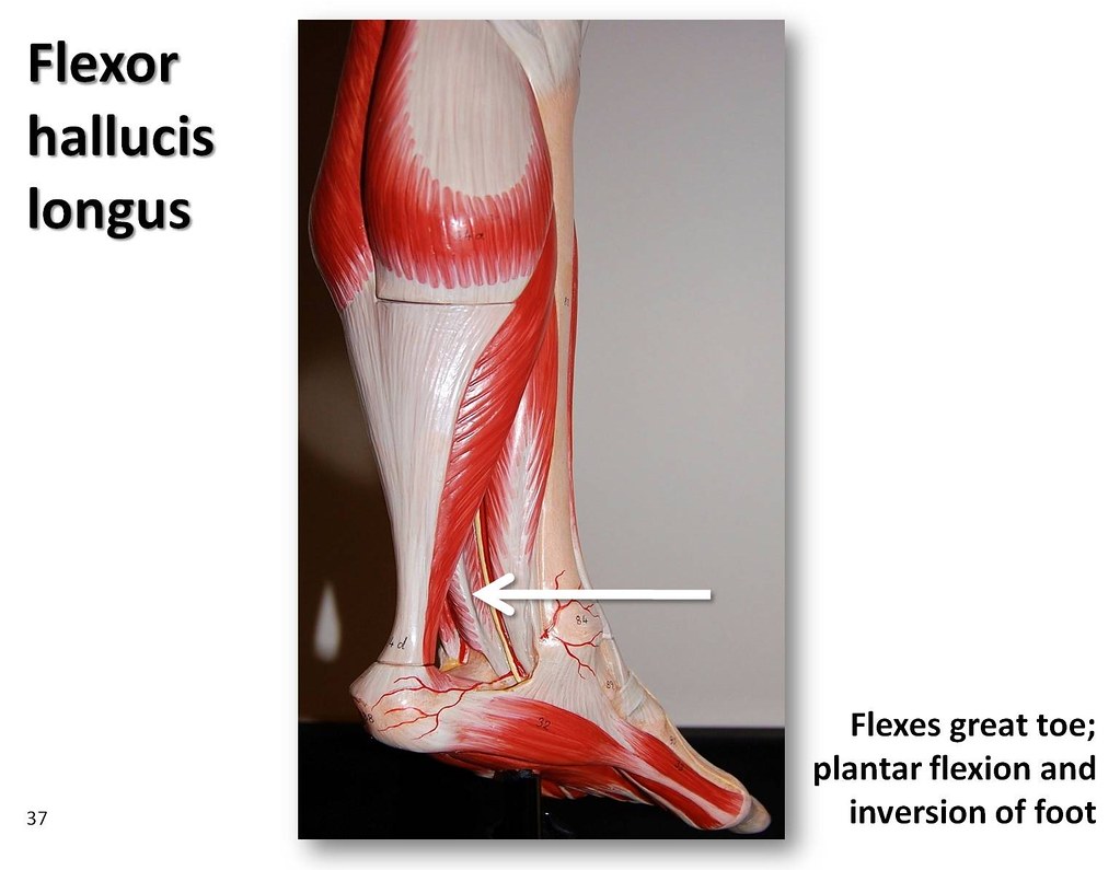
Flexor hallucis longus - Muscles of the Lower Extremity An ... from c1.staticflickr.com Localized anatomy of the hamstring muscles including semimembranosus, semitendinosus, biceps the hamstrings refer to 3 long posterior leg muscles, the biceps femoris, semitendinosus, and semimembranosus. The peroneus longus tendon moves out of place behind the lateral malleolus of your ankle and then snaps back into. Collectively, they act to dorsiflex and invert the foot at the ankle joint. 1280 x 1520 jpeg 166 кб. The pads of the machine are situated at the achilles tendon. Thompson's test, achilles tendon rupture. Related posts of muscle anatomy upper leg. Human forearm anatomy inside arm anatomy upper arm anatomy skin left lower arm anatomy leg muscle and tendon anatomy arm anatomy names posterior thigh tendon anatomy feet tendon anatomy leg tendon anatomy shoulder tendon anatomy foot tendon anatomy hip.
Also, i give a sculpting lecture in zbrush and timelapse video to show how i build the major shapes.
Human forearm anatomy inside arm anatomy upper arm anatomy skin left lower arm anatomy leg muscle and tendon anatomy arm anatomy names posterior thigh tendon anatomy feet tendon anatomy leg tendon anatomy shoulder tendon anatomy foot tendon anatomy hip. The human leg, in the general word sense, is the entire lower limb of the human body, including the foot, thigh and even the hip or gluteal region. The calcaneal tendon, also known as the tendon of achilles, is a posterior leg tendon — a fibrous connective tissue that joins muscles in the back of the leg. Thompson's test, achilles tendon rupture. Use the mouse scroll wheel to move the images up and down alternatively use the tiny arrows (>>) on both side of the image to move the images. Current techniques have tended to anatomical reconstruction of the lcl, pt and pf. Hands are outstretched, holding onto the handles of the bench. Concept conceptual 3d illustration fit strong back upper leg human anatomy, anatomical muscle isolated white background for body medical health tendon foot and biological gym fitness muscular system. Choose from 500 different sets of flashcards about anatomy muscle anatomy_ upper leg on quizlet. The thigh bone, or femur, is the large upper leg bone that connects the lower leg bones (knee joint) to the pelvic bone (hip joint). Related posts of muscle anatomy upper leg. Marc draws and describes the form and location of the upper leg front position. Tendons are fibrous cords attached to muscles and bone.


Belum ada Komentar untuk "Upper Leg Tendon Anatomy - Tushank Artworks - Page 2 : The pads of the machine are situated at the achilles tendon."
Posting Komentar